Fatty Liver and Estrogen Balance: Why Your Liver Health Is Key to Hormonal Harmony

You wake up tired even after a full night of sleep. The scale creeps up despite no real change in what you eat. Something feels off hormonally — but every test comes back “normal.” If any of this sounds familiar, it may not be random.
This may not be random — it may signal that your liver is struggling to keep up with its hormonal workload. And fatty liver and estrogen balance are far more tightly connected than most standard health conversations let on.
The encouraging news: understanding how fatty liver and estrogen balance interact is one of the most actionable things you can do for your metabolic health. Diet, movement, and a few specific lifestyle shifts can make a measurable difference — often within weeks.
What Is the Connection Between Fatty Liver and Estrogen Balance?

The liver is the body’s primary site for hormone clearance. It processes estrogen through a two-phase detoxification system, then packages it for elimination via bile and urine.
When excess fat accumulates in liver cells — defined as more than 5% fat by liver weight — this clearance process slows. The connection between fatty liver and estrogen balance becomes a two-way problem: impaired liver function disrupts hormone processing, and declining estrogen levels (particularly after menopause) remove a key metabolic protection, making fat accumulation more likely.[1]
Research shows that estrogen signaling through the ERα receptor actively protects liver cells from fat infiltration. Remove that signal — as happens naturally with age — and liver vulnerability increases significantly.
| Liver Function | Action | Health Outcome |
|---|---|---|
| Estrogen Clearance | Phase 1 + Phase 2 detox pathways break down and eliminate estrogen | Prevents estrogen buildup; supports hormonal equilibrium |
| ERα Receptor Signaling | Estrogen binds ERα in liver cells, activating protective pathways | Reduces hepatic fat accumulation |
| Transport Protein Production | Liver produces SHBG, which regulates active hormone levels in blood | Controls how much estrogen and testosterone are biologically active |
| Insulin Signal Processing | Healthy liver responds correctly to insulin, managing glucose output | Reduces fat storage cascade triggered by insulin resistance |
How the Liver Regulates Hormones — More Than Just a Filter
Most people think of the liver as a detox organ. That’s accurate but incomplete. It performs over 500 distinct metabolic tasks, and hormone regulation is one of the most consequential.
Hepatocytes — the specialized cells that make up roughly 70% of liver mass — are responsible for converting inactive hormone precursors into active forms, breaking down spent hormones, and producing the transport proteins that control how much of any given hormone is biologically available in the bloodstream at any moment.
Sex hormone-binding globulin (SHBG) is produced almost entirely in the liver. Low SHBG — a common marker in metabolic dysfunction — means more unbound estrogen and testosterone circulating freely. That has downstream effects on mood, energy, weight, and metabolic risk.
Roughly 60% of thyroid hormone conversion (T4 to active T3) also happens in the liver. This is why compromised liver function can produce symptoms that closely resemble hypothyroidism — fatigue, cold intolerance, brain fog — even when thyroid panels look normal on paper.[2]
When this system is working well, hormonal signals are processed efficiently and cleared on schedule. When the liver is burdened by fat accumulation, the entire cascade slows — and symptoms follow.
NAFLD: Causes, Stages, and What the Early Warning Signs Actually Feel Like
Non-alcoholic fatty liver disease (NAFLD) affects approximately one in four adults globally, making it the most common liver condition in the US. It’s defined by fat exceeding 5% of liver weight — in the absence of significant alcohol use.
What makes it particularly disruptive is how quietly it develops. Most people have no idea their liver is under strain until a routine blood panel catches elevated liver enzymes — or until symptoms have been present for years without a clear explanation.
Stages of Progression
| Stage | What’s Happening | Common Signs | Primary Risks |
|---|---|---|---|
| Simple Steatosis | Fat accumulation without significant inflammation | Often none; possible fatigue or upper-right abdominal discomfort | Progression to NASH; insulin resistance |
| NASH | Fat + inflammation + early cell damage | More pronounced fatigue, bloating, elevated liver enzymes | Fibrosis; accelerated metabolic decline |
| Fibrosis / Cirrhosis | Scarring replaces healthy tissue | Significant symptoms; can include jaundice, fluid retention | Liver failure; liver cancer |
Key risk factors include excess visceral fat, insulin resistance, high triglycerides, and sedentary behavior — the same cluster that defines metabolic syndrome.[3]
The early signs that often go unrecognized: persistent tiredness that doesn’t improve with rest, a dull ache in the upper right abdomen after large meals, and bloating that feels disproportionate to what was eaten. These aren’t dramatic. That’s exactly why they’re so frequently dismissed.
This cycle can develop quietly over years — which is why many people are genuinely caught off guard when a doctor first raises it. It is not a personal failure. It is a metabolic condition that responds well to targeted lifestyle changes, particularly when caught early.
Estrogen Detoxification: What Phase 1 and Phase 2 Actually Mean
The liver clears estrogen through a two-stage process. Understanding it helps explain why nutrient deficiencies — not just diet quality in general — can throw hormonal balance off in specific, predictable ways.
Phase 1 is handled primarily by cytochrome P450 enzymes. They convert estradiol into intermediate metabolites — some more potent, some less so. The balance between these intermediates matters: certain pathways produce more protective metabolites (2-hydroxyestrone), while others produce more proliferative ones (16α-hydroxyestrone).
Phase 2 attaches molecules to these intermediates — a process called conjugation — making them water-soluble so they can be excreted in bile or urine. This step depends heavily on specific nutrients: B vitamins (especially B6, B12, folate), magnesium, and sulfur-containing amino acids from foods like eggs, garlic, and cruciferous vegetables.[4]
If Phase 1 runs faster than Phase 2 can keep up — common when the liver is under metabolic stress — reactive intermediates accumulate. That imbalance is one mechanism behind symptoms like breast tenderness, mood shifts, and disrupted cycles in people who are still hormonally active.
Supporting Phase 2 is also why dietary fiber matters: it binds conjugated estrogen in the gut and escorts it out. Without adequate fiber, a gut enzyme called beta-glucuronidase can break that conjugation, allowing estrogen to be reabsorbed rather than eliminated.
Insulin Resistance and the Liver — A Two-Way Problem

Insulin resistance and liver fat don’t just coexist — they amplify each other. When cells stop responding properly to insulin, the pancreas compensates by producing more. Elevated insulin signals the liver to increase fat synthesis and storage — which accelerates fat accumulation in liver tissue.
A fat-burdened liver then releases more glucose into the bloodstream even in a fasted state, which triggers yet more insulin output. The cycle reinforces itself.[5]
For people with PCOS, this connection is particularly relevant. Chronically elevated insulin stimulates the ovaries to produce excess androgens — contributing to irregular cycles, acne, and metabolic disruption. Supporting liver function and insulin sensitivity addresses both sides of that equation simultaneously.
The same mechanism also explains why high-fructose intake is disproportionately damaging to liver health compared to other carbohydrates. Fructose is metabolized almost exclusively in the liver, where it can be converted directly into fat through a process called de novo lipogenesis — a connection that Harvard T.H. Chan School of Public Health research has highlighted in the context of sugar-sweetened beverage consumption.[6]
Most guides focus on “cutting carbs” as the primary lever here. That’s partly right — but it misses something. Fructose from whole fruit behaves differently than fructose from added sugars and sweetened beverages: the fiber, water content, and micronutrient context slow absorption and reduce the hepatic burden significantly. The issue isn’t fruit — it’s liquid fructose at high doses.
Why Risk Increases After Menopause — and What That Means Practically
Before menopause, estradiol actively protects liver cells from fat infiltration through ERα signaling. Circulating estradiol typically ranges from 30–400 pg/mL during reproductive years. After menopause, that drops to 0–30 pg/mL — removing a significant layer of metabolic protection.
Research suggests postmenopausal adults face approximately 2.4 times higher odds of developing NAFLD compared to premenopausal adults at similar metabolic baselines. The mechanism is direct: less ERα activation means less protective signaling against hepatic fat accumulation.[1]
| Life Stage | Estradiol Range | Hepatic Risk Level | Key Protective Factor |
|---|---|---|---|
| Premenopausal | 30–400 pg/mL | Lower | Active ERα signaling; higher SHBG |
| Perimenopausal | Fluctuating | Transitional — rising | Lifestyle intervention has highest impact here |
| Postmenopausal | 0–30 pg/mL | Higher (~2.4×) | Diet, exercise, sleep quality, toxin reduction |
| Aging Males | Testosterone declining | Moderate, increasing | Maintaining healthy androgen-to-estrogen ratio |
This is also where the perimenopause window is underappreciated. Hormonal fluctuation — not just the eventual decline — already begins to shift metabolic risk. Lifestyle intervention during the perimenopause transition may offer more leverage than intervention that starts after the fact.
What the ERα Research Actually Shows
Much of what we know about estrogen’s liver-protective role comes from mouse models — specifically αERKO and KIKO models, which allow researchers to isolate the exact role of the ERα receptor.
αERKO mice lack the ERα receptor entirely. On high-fat diets, they develop significantly greater microvesicular steatosis than wild-type counterparts — meaning more fat droplets accumulating inside liver cells.
KIKO mice retain the receptor but cannot bind DNA directly. They show intermediate fat accumulation, confirming that direct DNA binding of ERα — not just its presence — is required for full hepatoprotective effect.
The takeaway isn’t that humans should be supplementing estrogen (that’s a clinical conversation requiring individual assessment). The research insight is mechanistic: estrogen doesn’t just affect reproductive tissue — it actively shapes metabolic function in the liver at a cellular level, and its decline has real, measurable consequences for hepatic fat accumulation.[1]
| Mouse Model | ERα Status | Observed Effect | Insight |
|---|---|---|---|
| Wild-Type | Normal function | Minimal fat accumulation | Baseline: healthy ERα signaling protects liver |
| αERKO | Receptor absent | High microvesicular steatosis | ERα is the primary protective mediator |
| KIKO | Present but DNA binding impaired | Intermediate fat infiltration | Direct DNA binding — not just receptor presence — is required |
The Gut-Liver Axis — Where Microbiome Meets Hormonal Health
The liver and gut are physically connected via the portal vein, which carries nutrients and microbial metabolites directly from the intestines to hepatic tissue. This means gut health is never separate from liver health — it’s upstream of it.
Inside the gut lives a specialized subset of bacteria called the estrobolome — bacteria that produce an enzyme called beta-glucuronidase. In a healthy gut, beta-glucuronidase activity is balanced: it helps recycle some estrogen metabolites while allowing others to be excreted.
When the microbiome becomes imbalanced — a state called dysbiosis, linked to low-fiber diets, chronic stress, antibiotics, and poor sleep — beta-glucuronidase activity can rise. That leads to more reactivated estrogen re-entering circulation rather than being eliminated. The result is a higher effective estrogen burden on the liver, which compounds the clearance problem.[7]
Constipation makes this worse. The longer conjugated hormones sit in the colon, the more opportunity there is for reabsorption. This is one reason why bowel regularity — not just diet composition — is a meaningful variable in hormonal health.
Dietary fiber feeds the beneficial bacteria that keep beta-glucuronidase in check. Prebiotic foods like garlic, leeks, onions, and Jerusalem artichokes are particularly effective. Fermented foods like plain yogurt, kefir, and kimchi support microbial diversity more broadly.
How to Support Fatty Liver and Estrogen Balance Through Lifestyle
This is the section where most guides list five generic habits and call it done. The actual picture is more specific — and more actionable — than that.
Nutrition: What Matters Most
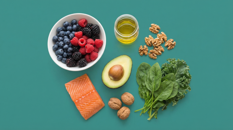
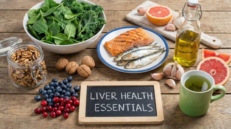
Cruciferous vegetables — broccoli, kale, Brussels sprouts, cauliflower, arugula — contain a compound called indole-3-carbinol that actively supports Phase 2 estrogen detox and has been studied for its role in shifting estrogen metabolism toward more protective pathways. These aren’t interchangeable with “vegetables in general.”
Ground flaxseed (2–3 tablespoons daily) provides both lignans (phytoestrogens with mild estrogen-modulating effects) and soluble fiber. Fiber intake of 25–35 grams daily supports estrogen clearance via the gut. Most adults in the US average closer to 15 grams — leaving a meaningful gap.
Quality protein — eggs, fatty fish like salmon and sardines, Greek yogurt, legumes — provides the amino acids required for Phase 2 conjugation reactions. Skipping protein doesn’t just affect satiety; it directly limits the liver’s detox capacity.
Olive oil, avocado, and walnuts supply healthy fats that reduce liver inflammation and support bile production — which is how conjugated hormones are actually exported from the liver. Alcohol, added fructose (from sweetened beverages, not whole fruit), and ultra-processed foods work against all of these pathways simultaneously.
For a deeper breakdown of which foods support hepatic function specifically, see the best foods for liver health guide on this site.

Movement: Timing and Type Both Matter
150 minutes of moderate movement per week — roughly 20–25 minutes daily — is the threshold supported by evidence for meaningful metabolic benefit. Even short sessions after meals may help reduce post-meal blood sugar spikes, which directly reduces insulin load on the liver.
Resistance training deserves particular attention here. Skeletal muscle is the body’s largest glucose sink — accounting for approximately 80% of insulin-mediated glucose uptake according to the American Diabetes Association. Building and maintaining muscle mass reduces the amount of glucose that would otherwise be processed (and potentially stored as fat) by the liver.
Sleep and Stress: The Variables Most Plans Ignore
One thing worth pushing back on here: the standard advice treats nutrition and exercise as the primary levers for liver and hormonal health. But research published in the Journal of Applied Physiology found that just one week of sleep restriction (to about 5 hours per night) reduced insulin sensitivity by 30–40%. That’s a larger acute effect than most dietary interventions produce.
Cortisol — the primary stress hormone — is also processed by the liver. Chronic stress elevates cortisol, which drives visceral fat accumulation and increases hepatic fat storage independently of diet. Managing stress isn’t a soft recommendation — it’s a direct metabolic input.
Seven to nine hours of quality sleep, alongside stress-reduction practices like consistent meal timing, outdoor movement, and breathing work, supports hormone clearance efficiency in ways that no dietary supplement can replicate.
Reducing Environmental Estrogen Load
Endocrine-disrupting chemicals — xenoestrogens found in plastics (particularly BPA and phthalates), pesticide residues, and synthetic fragrance — compete with natural estrogen for receptor binding and add to the liver’s detox burden. Reducing exposure through practical swaps can meaningfully lighten that load:
- Store food and beverages in glass or stainless steel rather than plastic, especially for acidic or fatty foods
- Choose fragrance-free personal care products or those with disclosed ingredient lists
- Prioritize the “dirty dozen” produce list for organic purchasing; washing all produce thoroughly also helps
- Avoid heating food in plastic containers
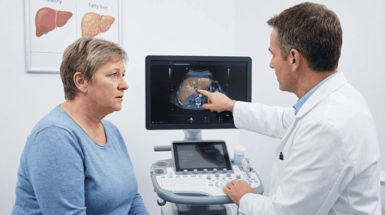
Understanding liver enzyme markers — and what elevated ALT or AST actually indicates — can help track whether these interventions are working over time.
Conclusion
The connection between fatty liver and estrogen balance isn’t a niche finding. It’s a clinically relevant, mechanistically documented relationship that sits at the intersection of hormonal health, metabolic function, and aging.
What the research shows, practically: estrogen protects the liver from fat accumulation through ERα signaling; a burdened liver, in turn, processes hormones less efficiently — creating a feedback loop that compounds over time. Add insulin resistance and gut dysbiosis to the picture, and you have a system under strain from multiple directions simultaneously.
The leverage points are real and accessible. Cruciferous vegetables and adequate fiber support Phase 2 detox and gut estrogen clearance. Resistance training and quality sleep may do more for insulin sensitivity than most dietary changes. Reducing xenoestrogen exposure lightens the liver’s detox burden.
These aren’t dramatic interventions. They’re consistent, targeted ones — and that’s what the evidence actually supports. You don’t need a perfect protocol. You need a sustainable one that addresses the root mechanisms.
If you suspect liver function may be contributing to the symptoms you’re experiencing, the fatty liver disease overview on this site is a good next read — and a conversation with your doctor about liver enzyme panels is a reasonable first clinical step.
Frequently Asked Questions
What is the connection between fatty liver and estrogen balance?
Fatty liver and estrogen balance are linked in two directions. A healthy liver processes and clears estrogen through Phase 1 and Phase 2 detoxification pathways — when liver function is impaired by fat accumulation, estrogen clearance slows, which can disrupt hormonal equilibrium. In the other direction, estrogen itself — acting through ERα receptors in liver cells — actively protects against fat buildup. When estrogen levels decline (as they do with aging and menopause), that protective signaling weakens, making the liver more susceptible to fat accumulation. Research suggests this bidirectional relationship is a key driver of why metabolic risk increases during hormonal transitions.
What are the early signs that liver function may be affecting hormone balance?
Early signs are often subtle and easy to attribute to other causes: persistent fatigue that sleep doesn’t resolve, unexplained weight gain particularly around the midsection, bloating after meals, and mood fluctuations. Because the liver produces sex hormone-binding globulin (SHBG), impaired liver function can also shift the ratio of active to bound hormones — contributing to symptoms like low libido or irregular cycles in people who are still hormonally active. If a standard thyroid panel comes back normal but symptoms persist, liver function (specifically ALT and AST enzyme levels) is worth investigating with your doctor.
Why does NAFLD risk increase after menopause?
Before menopause, estradiol actively protects liver cells by activating ERα receptors, which suppress fat accumulation pathways. As estradiol levels drop — from a range of 30–400 pg/mL to 0–30 pg/mL — that protective signaling diminishes. Research suggests postmenopausal adults face roughly 2.4 times higher odds of developing NAFLD compared to premenopausal adults at similar metabolic baselines. The perimenopause transition is also a high-leverage window: beginning lifestyle interventions before the full hormonal shift may provide more protection than waiting until after.
How are insulin resistance and liver fat connected?
They reinforce each other in a self-amplifying cycle. Insulin resistance causes the pancreas to overproduce insulin; elevated insulin signals the liver to increase fat synthesis and storage. A fat-burdened liver then releases excess glucose even when fasting, which drives more insulin output. Supporting insulin sensitivity — through resistance training, adequate sleep, reduced added sugar intake, and regular movement — directly reduces the hepatic fat accumulation that perpetuates the cycle. Addressing one side of this equation consistently improves the other.
Which dietary changes have the most direct impact on liver and estrogen health?
The evidence points most clearly toward three areas: increasing cruciferous vegetables (broccoli, kale, Brussels sprouts) for Phase 2 detox support; reaching 25–35 grams of dietary fiber daily to support estrogen clearance through the gut; and prioritizing quality protein at each meal (eggs, fatty fish, legumes, Greek yogurt) to supply amino acids required for conjugation reactions. Reducing added fructose from sweetened beverages — not whole fruit — specifically targets hepatic de novo lipogenesis. These aren’t replacements for each other; the combination is what supports the full detox and clearance pathway.
Medical Disclaimer: The information provided in this article is for educational purposes only and does not constitute medical advice. Always consult a qualified healthcare provider before making changes to your diet, lifestyle, or treatment plan. TheMetabolicHub.com does not replace professional medical guidance.
References
- Zhu L, Brown WC, Cai Q, et al. Estrogen treatment after ovariectomy protects against fatty liver and may improve pathway-selective insulin resistance. Diabetes. 2013;62(2):424–434. PMID: 23043160
- Pucci E, Chiovato L, Pinchera A. Thyroid and lipid metabolism. Int J Obes Relat Metab Disord. 2000;24 Suppl 2:S109–S112. PMID: 10997617
- Chalasani N, Younossi Z, Lavine JE, et al. The diagnosis and management of nonalcoholic fatty liver disease. Hepatology. 2018;67(1):328–357. PMID: 28714183 — bitte vor Publikation verifizieren
- Hodges RE, Minich DM. Modulation of metabolic detoxification pathways using foods and food-derived components. J Nutr Metab. 2015;2015:760689. PMC: PMC10296738
- American Diabetes Association. Insulin resistance and type 2 diabetes. diabetes.org
- Harvard T.H. Chan School of Public Health. The Nutrition Source — Fructose and liver fat. hsph.harvard.edu
- Baker JM, Al-Nakkash L, Herbst-Kralovetz MM. Estrogen-gut microbiome axis: Physiological and clinical implications. Maturitas. 2017;103:45–53. PMID: 28778332